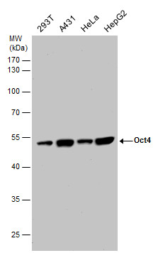
GTX100622 WB Image
OCT4 antibody detects OCT4 protein by western blot analysis. Various whole cell extracts (30 ug) were separated by 10% SDS-PAGE, and the membrane was blotted with OCT4 antibody (GTX100622) diluted at a dilution of 1:10000. The HRP-conjugated anti-rabbit IgG antibody (GTX213110-01) was used to detect the primary antibody.
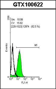
GTX100622 FACS Image
Flow cytometry on human embryonic stem cells, staining with Oct4 (GTX100622)antibody at 1:50 dilution(green) or rabbit IgG (black).
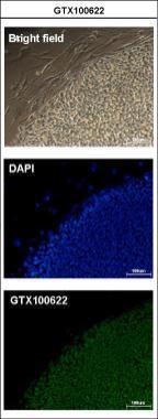
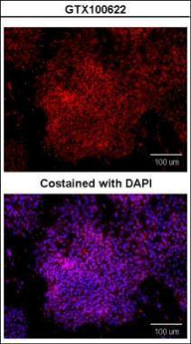
GTX100622 ICC/IF Image
Immunofluorescence analysis of paraformaldehyde-fixed human embryonic stem cell, using Oct4(GTX100622) antibody at 1:200 dilution.
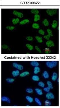
GTX100622 ICC/IF Image
Immunofluorescence analysis of paraformaldehyde-fixed Human ESC, using Oct4(GTX100622) antibody at 1:400 dilution.
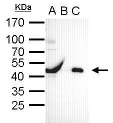
GTX100622 IP Image
Oct4 antibody immunoprecipitates Oct4 protein in IP experiments. IP Sample: cell lysate/extract of Oct4 gene transfected 293T cells A. Cell lysate/extract of transfected 293T cell B. Control with 2 ug of preimmune rabbit IgG C. Immunoprecipitation of Oct4 by 2 ug of Oct4 antibody (GTX100622) 12% SDS-PAGE The immunoprecipitated Oct4 protein was detected by Oct4 antibody (GTX100622) diluted at 1:1000. EasyBlot anti-rabbit IgG (GTX221666-01) was used as a secondary reagent.
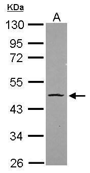
GTX100622 WB Image
Sample (20 ug)
A: HeLa nucleus
10% SDS PAGE
GTX100622 diluted at 1:1000
The HRP-conjugated anti-rabbit IgG antibody (GTX213110-01) was used to detect the primary antibody.
GTX100622 ICC/IF Image
Immunofluorescence analysis of paraformaldehyde-fixed human embryonic stem cell, using Oct4(GTX100622) antibody at 1:100 dilution.
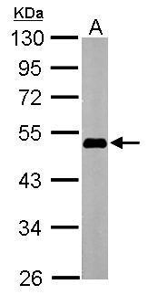
GTX100622 WB Image
Sample (20 ug of whole cell lysate)
A: Human ESC
10% SDS PAGE
GTX100622 diluted at 1:1000
The HRP-conjugated anti-rabbit IgG antibody (GTX213110-01) was used to detect the primary antibody.

GTX100622 WB Image
Sample (20 ug of whole cell lysate)
A: mouse ESC
12% SDS PAGE
GTX100622 diluted at 1:5000
The HRP-conjugated anti-rabbit IgG antibody (GTX213110-01) was used to detect the primary antibody.